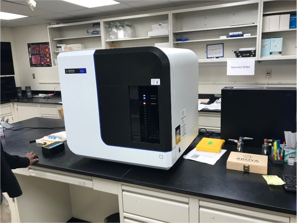
The Vectra Polaris scans both brightfield and fluorescent slides and can take multispectral imagery of user-specified areas of interest, or user-chosen TMA cores. The whole slide multispectral imaging capability creates a simpler and more robust workflow as fields of view do not need to be selected eliminating selection bias. The Vectra Polaris is equipped with a 10x, 20x, and 40x objective, as well as a liquid crystal tunable filter that generates unmixed, annotated regions of interest of up to 9-colors for deeper interrogation of biology. After imaging is complete you retain a whole slide record, no re-scans required, so that you can easily re- analyze imagery as new understanding emerges. Tissue sections or TMAs can be labeled with immunofluorescent (IF) or immunohistochemical (IHC) stains such as Opal™, or with conventional stains such as H&E and trichrome. When using IF or IHC stains, multiple proteins can be measured on a per tissue, per cell, or per cell compartment. Enclosed system with built-in touchless automation allows you to visualize, analyze, quantify and phenotype immune cells in situ with enhanced security and reliability. Integrated inForm and phenoptr tissue analysis software packages support configurable projects for biomarker quantification and enhanced spatial analysis with machine learning.
For Training and use of this equipment contact: Mercedes Gonzalez-Juarrero (970-491-7306; Mercedes.gonzalez-juarrero@colostate.edu)
Cost:
Training: $250 including 5 h of use.
Hourly: $20/h for first 4 hr, $3/hr thereafter.

