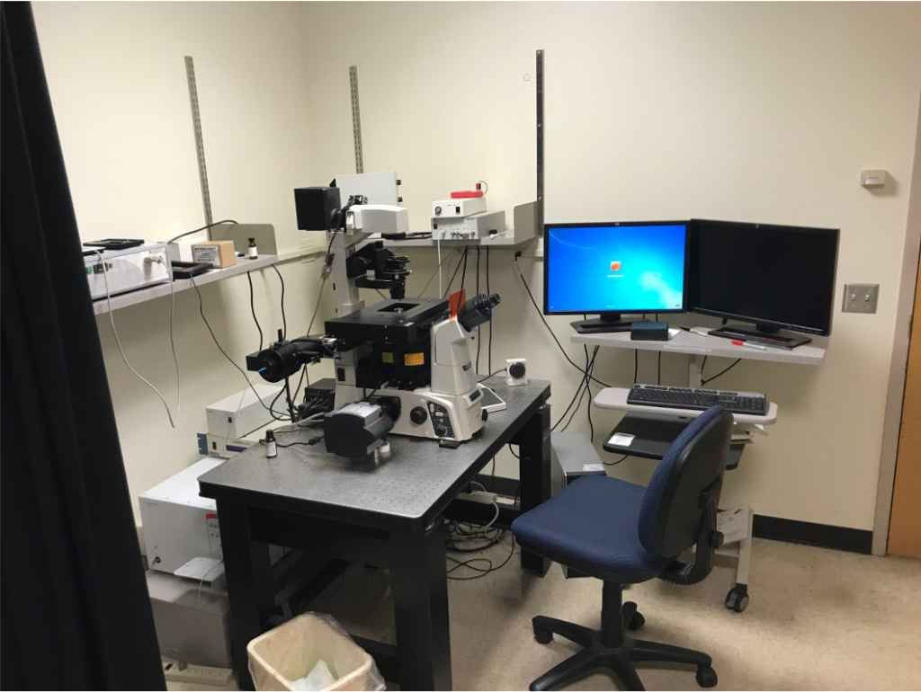
Location: Room 224 MRB
The Nikon Total Internal Reflection Fluorescence (TIRF) Inverted Microscope (Eclipse Ti) has perfect focus control, a four laser-line launch (405, 488, 561 and 640 nm) and an Intensilight Hg illumination source. A motorized XY Piezo Z stage and stage-top environmental chamber with temperature, CO2/air, humidity capability, will support culture dishes, multi-well plates or slides. Equipped with 20X, 40X, 60X DIC objectives and a 100X TIRF objective. Microscope has an Andor Clara camera for wide field imaging and an Andor iXon3 EMCCD camera for TIRF imaging. Microscope has built-in N-STORM super-resolution imaging capability and is networked to a RAID array for multiple terabyte storage. Nikon elements software controls all aspects of acquisition and analysis with a deconvolution software package included. NIS-Elements Software is the central tool required for multi-dimensional and time-lapse imaging. Galvo-based point scanner. Miniscanner unit with one set of 3 mmgalvos, custom tuned for high-speed precision pointing.
For Training and use of this equipment contact: Keith DeLuca, 970-491-5149 (keith.deluca@colostate.edu)
Cost:
Training: $250 includes 5 h of use.
Hourly: $20 first hr; $3/hr thereafter
Top left: A conventional fluorescence microscopy image of all the telomeres in a cell. Bottom left: A map of fluorescent probes detected by the STORM microscope, overlaying a single telomere from the conventional image. Right: the same map of probes from an individual telomere, enlarged and in three dimensions. Credit: Chris Nelson/Bailey Lab. (Image reproduced from https://source.colostate.edu/exquisite-resolution-microscopes-illuminate-hidden-intracellular-worlds/)

