Bruker Atomic Force Microscope
Location: Room 362 Scott Bioengineering Building
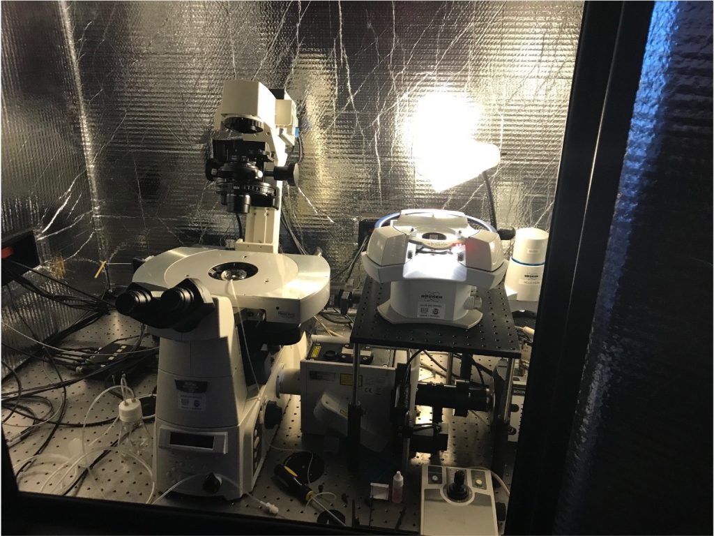
This Bruker BioScope Resolve AFM System can be used alone or integrated with an inverted light microscopes (see separate entry for Nikon Eclipse Spinning Disk Confocal Microscope). It is configured to provide the industry standard highest resolution imaging in both air and fluid samples in contact mode, tapping mode, and Bruker’s unique PeakForce Tapping mode. The cantilever deflection is detected with an IR (850 nm) laser to prevent interference with optical imaging modes. Air and fluid cantilever holders are included. Standard magnetic sample clamps for slides, coverslips, petri dishes, and contact and tapping mode tips are also included for calibration and training.
The Bruker’s Premium Accessory Package for the Resolve includes a sample heater accessory for heating the stage to physiological temperature (or higher). Combined with the Petri Dish Perfusion Accessory, this will enable live cell imaging and even long-term maintenance of cell cultures on the microscope stage, with both liquid and gas ports to maintain the culture environment. This package also includes the Nanomechanics package and the Microscope Image Registration & Overlay (MIRO) software package. The Nanomechanics package provides quantitative force volume mapping for high-resolution nanomechanical characterization. The MIRO package automates importing images from the optical microscope in real-time to guide the AFM imaging and force measurements to regions of interest identified in an optical image, for example. When performing AFM imaging, fluorescence micrographs can be taken simultaneously after every AFM line scan.
The NanoMan Software provides flexible, accurate control of the of the AFM probe for nanomanipulation and nanolithography applications, using either graphical “point-and-click” and script-driven modes. This also includes the NanoScope Analysis Software for image and data (e.g. force curve) analysis.
Acoustic Enclosure and Vibration Isolation. Herzan’s AEK-2011 highly attenuating acoustic enclosure with broad access is perfectly suited for AFM. This enclosure will provide high-performance acoustic isolation. The vibration isolation system TS-300LT provides active isolation in all six degrees of freedom from 0.7 to 1000 Hz, and passive isolation above 1000 Hz.
For Training and use of this equipment contact: Ellen Brennan-Pierce, 970-491-5046 (ellen.brennan-pierce@colostate.edu)
Cost (Pending BFS approval):
Training: $250 includes 5 h of use.
AFM (with or without SDC): $45 for first hour and $5/hr thereafter
Nikon Eclipse Inverted Confocal Microscope
Location: 362 Scott Biomedical Engineering
The Nikon Eclipse Inverted Confocal has perfect focus control and a four laser-line launch (405, 488, 561 and 640 nm) and an Intensilight Hg illumination source. A motorized XY Piezo Z stage and stage-top with temperature control will support culture dishes, multi-well plates or slides. Equipped with planapo/DIC 10x, 20X, 40X, and 100X (oil) objectives. Nikon elements software controls all aspects of acquisition and analysis with a deconvolution software package included. The software is the central tool required for multi-dimensional and time-lapse imaging.
This microscope is also used in conjunction with the Bruker AFM (described elsewhere) for applications to cell or cell-substrate imaging.
Key components include:
1. Motorized inverted microscope Nikon Eclipse Ti-E equipped with extra fine/fine/coarse focusing drive, motorized nosepiece, motorized filter cube turret, motorized x,y stage, DIC components, CFI plan apochromat lambda 10x, 20x, 40x, and 100x oil immersion objectives.
2. LUNV monolithic laser combiner with and acousto-optic tunable filter (AOTF) with 20 mW 405 nm, 70 mW 488 nm, 70 mW 561 nm, and 40 mW 640 nm lines.
5. Galvo-based point scanner. Miniscanner unit with one set of 3 mmgalvos, custom tuned for high-speed precision pointing.
6. Yokogawa CSU-X1 Spinning Disk Unit. This is the most widely used solution in confocal cell imaging for its high speed, low phototoxicity, and extremely low noise.
7. Andor iXon DU897 Ultra EMCCD camera. This thermoelectrically-cooled camera allows for single molecule sensitivity at 56 fps (full frame) in fluorescence measurements.
For Training and use of this microscope contact: Ellen Brennan-Pierce (TEL 970-491-5046; email: ellen.brennan-pierce@colostate.edu)
Cost (Pending BFS approval):
Training: $250 per session
Use: $20 for first hr; $3/hr thereafter.